Acute Occlusion Of Middle Cerebral Artery Radiology F Vrogue Co

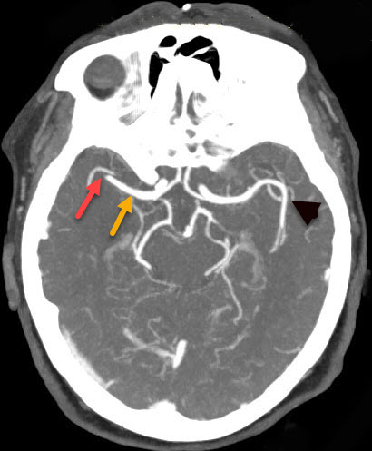
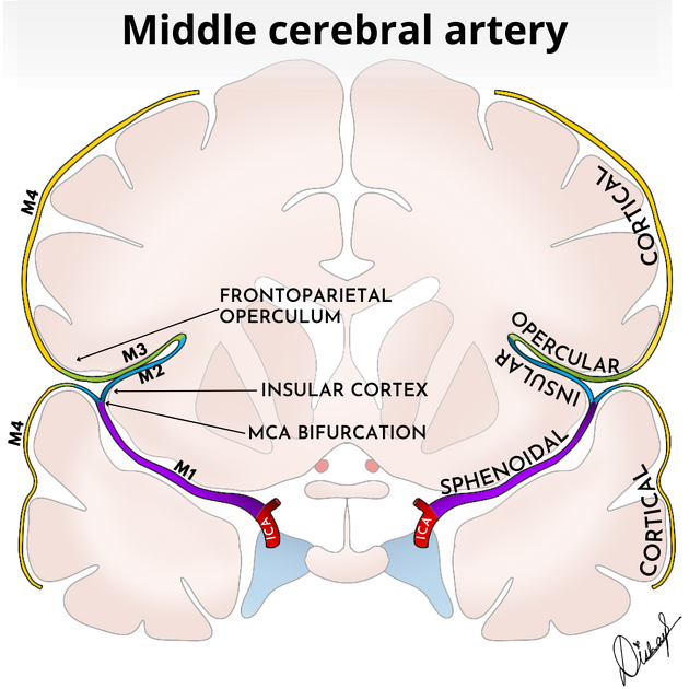
Mca Occlusion Stroke Mca Segments Radiology Radiology For Beginners The patient underwent catheter digital subtraction angiography, which confirmed acute left middle cerebral artery m1 segment occlusion. aspiration thrombectomy was performed, achieving tici 3 reperfusion following a single pass. Learn about mca occlusion stroke, radiology of mca segments, and cerebral artery occlusion. visit now for expert tips and insights!.

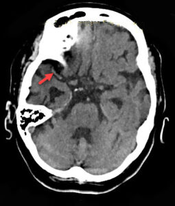
Mca Occlusion Stroke Mca Segments Radiology Radiology For Beginners Here, we review the focal cerebral ischemia models with an emphasis on the middle cerebral artery occlusion technique, a “gold standard” in surgical ischemic stroke models. This chapter discusses the diagnosis and treatment of acute ischemic stroke from large vessel occlusion, including the rapid radiographic evaluation with ct, cta, mri, and perfusion imaging. There are however certain features specific to middle cerebral artery infarct, and these are discussed below. for both ct and mri it is worth dividing the features according to the time course. Background and purpose— we sought to evaluate how accurately length and volume of thrombotic clots occluding cerebral arteries of patients with acute ischemic stroke can be assessed from nonenhanced ct (nect) scans reconstructed with different slice widths.

Acute Occlusion Of Middle Cerebral Artery Radiology F Vrogue Co There are however certain features specific to middle cerebral artery infarct, and these are discussed below. for both ct and mri it is worth dividing the features according to the time course. Background and purpose— we sought to evaluate how accurately length and volume of thrombotic clots occluding cerebral arteries of patients with acute ischemic stroke can be assessed from nonenhanced ct (nect) scans reconstructed with different slice widths. In the present study, patients with acute middle cerebral artery occlusion (mcao) underwent diffusion weighted imaging (dwi) to evaluate whether the location of the infarct core could be used to differentiate icas from intracranial embolism. The mri findings and clinical symptoms are consistent with acute cerebral infarction due to middle cerebral artery stenosis caused by vasculitis (possible central nervous system vasculitis). The clinical outcome of 40 cases with middle cerebral artery (mca) occlusion was examined in relation to the site of occlusion and the findings on computed tomography (ct). Till now, studies investigating the use of ctp in distinguishing icas from embolism as the cause of acute mcao are still scarce. hence, the present study was conducted to evaluate the effectiveness of the baseline ctp imaging characteristics in identifying icas related acute mcao.

Acute Occlusion Of Middle Cerebral Artery Radiology F Vrogue Co In the present study, patients with acute middle cerebral artery occlusion (mcao) underwent diffusion weighted imaging (dwi) to evaluate whether the location of the infarct core could be used to differentiate icas from intracranial embolism. The mri findings and clinical symptoms are consistent with acute cerebral infarction due to middle cerebral artery stenosis caused by vasculitis (possible central nervous system vasculitis). The clinical outcome of 40 cases with middle cerebral artery (mca) occlusion was examined in relation to the site of occlusion and the findings on computed tomography (ct). Till now, studies investigating the use of ctp in distinguishing icas from embolism as the cause of acute mcao are still scarce. hence, the present study was conducted to evaluate the effectiveness of the baseline ctp imaging characteristics in identifying icas related acute mcao.

Acute Occlusion Of Middle Cerebral Artery Radiology F Vrogue Co The clinical outcome of 40 cases with middle cerebral artery (mca) occlusion was examined in relation to the site of occlusion and the findings on computed tomography (ct). Till now, studies investigating the use of ctp in distinguishing icas from embolism as the cause of acute mcao are still scarce. hence, the present study was conducted to evaluate the effectiveness of the baseline ctp imaging characteristics in identifying icas related acute mcao.

Comments are closed.