Anatomy Of The Heart

Heart Anatomy Vector Illustration Stock Vector Colourbox Your heart contains four muscular sections (chambers) that briefly hold blood before moving it. electrical impulses make your heart beat, moving blood through these chambers. This page discusses the heart anatomy. click here to learn everything about the anatomy of the heart, heart valves and vessels at kenhub!.

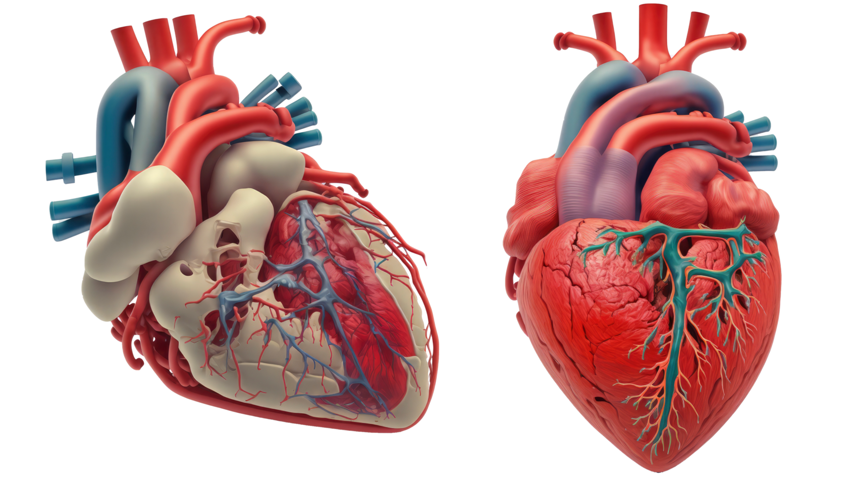
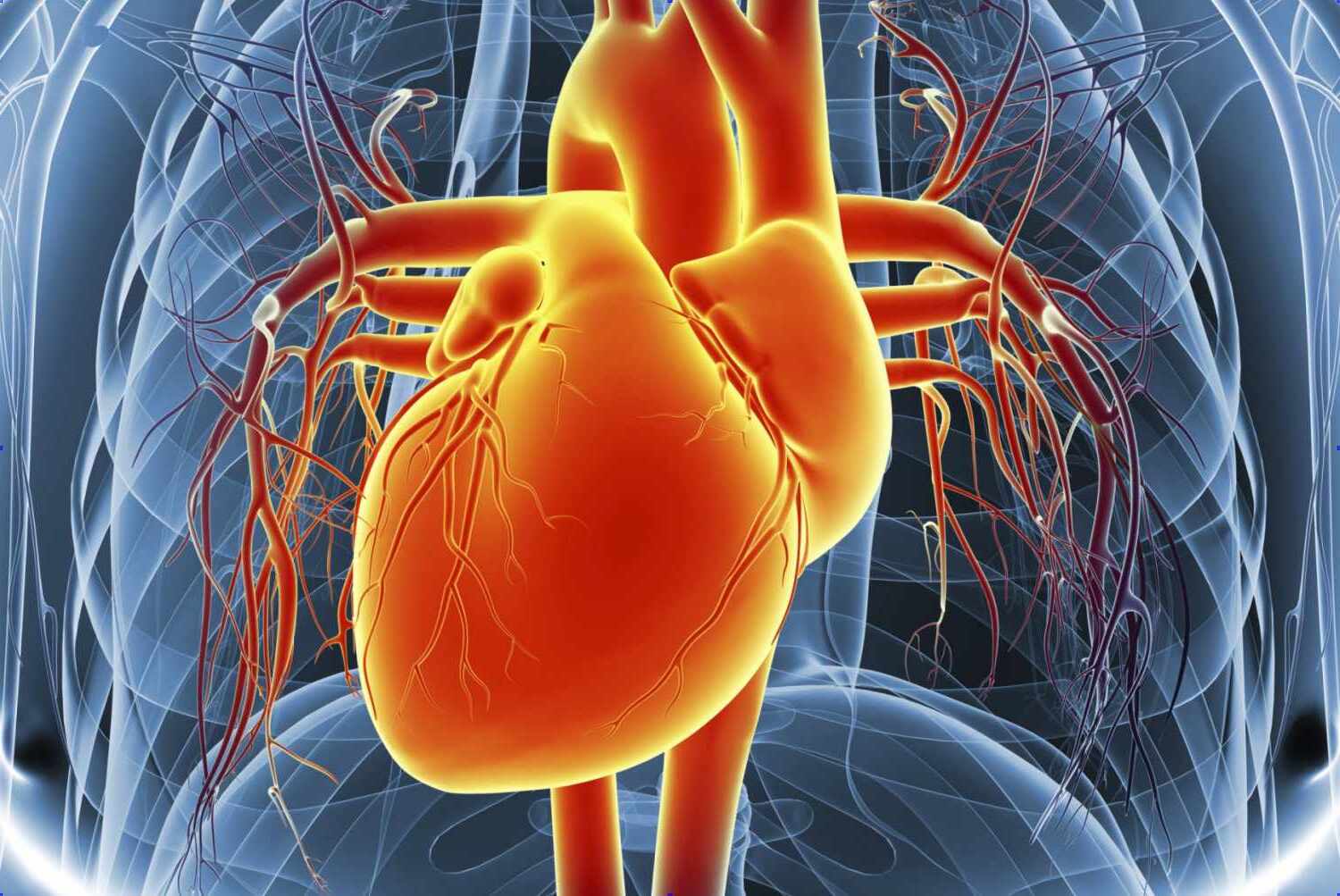
Anatomy Heart Png File The heart consists of several layers of a tough muscular wall, the myocardium. a thin layer of tissue, the pericardium, covers the outside, and another layer, the endocardium, lines the inside. This detailed anatomical illustration showcases the heart's major vessels, arteries, and veins, along with its essential structural components, providing a comprehensive view of cardiac anatomy from an anterior perspective. Explore the heart anatomy with its parts, names & a clear diagram. learn how the heart functions & its crucial role in the circulatory system. Blood flows from the body and lungs to the atria and from the atria to the ventricles. the ventricles pump blood out of the heart to the lungs and other parts of the body. an internal wall of tissue divides the right and left sides of your heart. this wall is called the septum.

Heart Anatomy Quiz Doquizzes Explore the heart anatomy with its parts, names & a clear diagram. learn how the heart functions & its crucial role in the circulatory system. Blood flows from the body and lungs to the atria and from the atria to the ventricles. the ventricles pump blood out of the heart to the lungs and other parts of the body. an internal wall of tissue divides the right and left sides of your heart. this wall is called the septum. The heart is a muscular organ situated in the mediastinum. it consists of four chambers, four valves, two main arteries (the coronary arteries), and the conduction system. Learn about the heart's anatomy, how it functions, blood flow through the heart and lungs, its location, artery appearance, and how it beats. It consists of four main chambers: two atria and two ventricles. 1 understanding its basic anatomy is crucial to understanding how it functions. this article provides a comprehensive look at the heart's structure with a detailed, labeled diagram and realistic photos, guiding you through each part and its role in the circulatory system. In cardiac anatomy, knowledge of the relative disposition of the cardiac chambers is as relevant as the intrinsic chamber morphology. this review considers the cardiac chambers, coronary arteries and the conduction system.

Queensland Cardiovascular Group Anatomy Of The Heart The heart is a muscular organ situated in the mediastinum. it consists of four chambers, four valves, two main arteries (the coronary arteries), and the conduction system. Learn about the heart's anatomy, how it functions, blood flow through the heart and lungs, its location, artery appearance, and how it beats. It consists of four main chambers: two atria and two ventricles. 1 understanding its basic anatomy is crucial to understanding how it functions. this article provides a comprehensive look at the heart's structure with a detailed, labeled diagram and realistic photos, guiding you through each part and its role in the circulatory system. In cardiac anatomy, knowledge of the relative disposition of the cardiac chambers is as relevant as the intrinsic chamber morphology. this review considers the cardiac chambers, coronary arteries and the conduction system.

Comments are closed.