Aortic Dissection On Chest X Ray

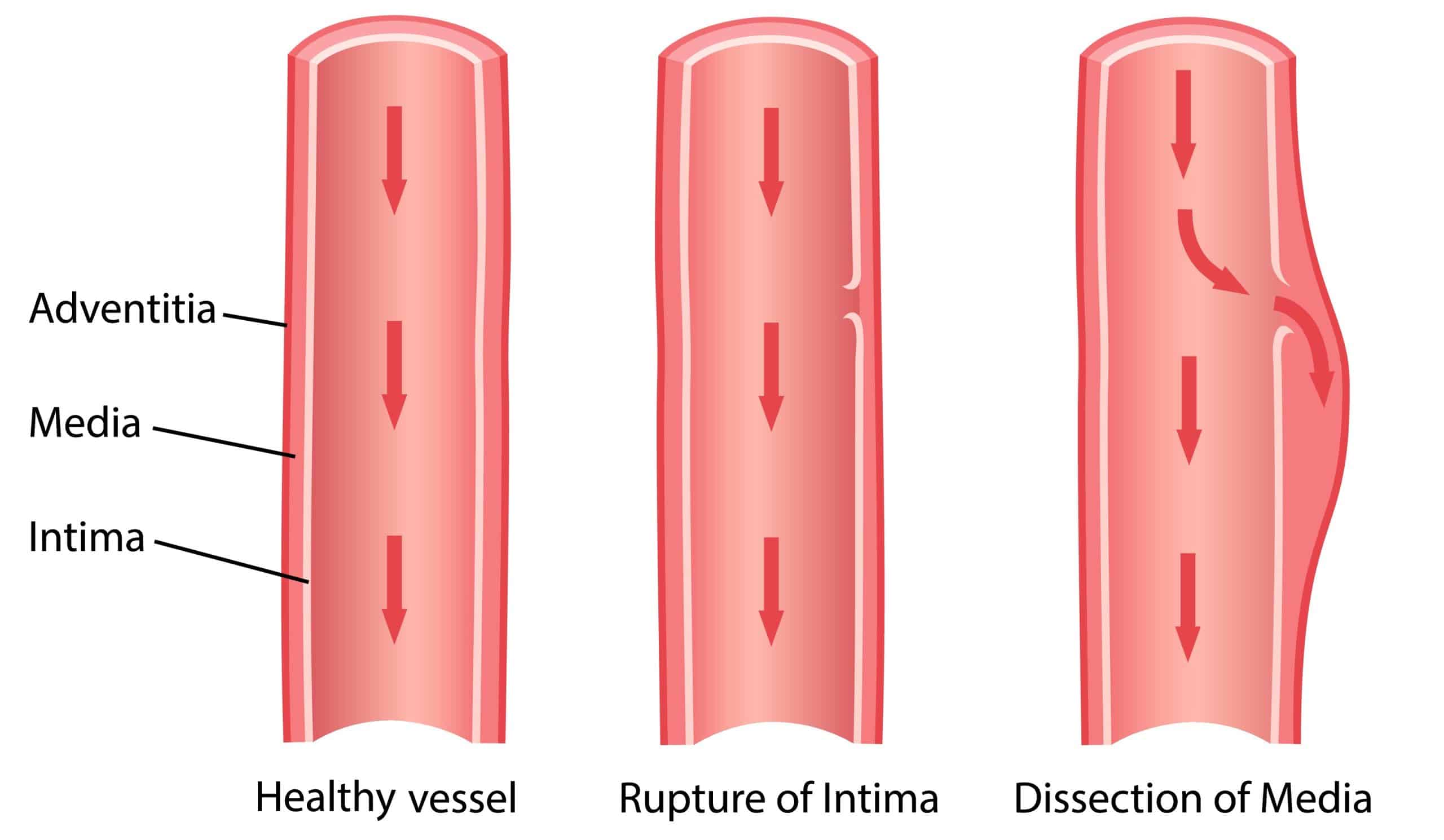
Aortic Dissection Aortic Dissection Chest X Ray Image Vrogue Co Aortic dissection is the prototype and most common form of acute aortic syndromes and a type of arterial dissection. it occurs when blood enters the medial layer of the aortic wall through a tear or penetrating ulcer in the intima and tracks longitudinally along with the media, forming a second blood filled channel (false lumen) within the. Chest x ray may be helpful in the diagnosis of aortic dissection. findings suggestive of aortic dissection on x ray include widening of mediastinum, wide aortic contour, tracheal deviation, aortic kinking, and displacement of previous aortic calcification.

Aortic Dissection Aortic Dissection Chest X Ray Image Vrogue Co Learn how chest x rays assist in identifying aortic dissection by highlighting key radiographic indicators and differentiating it from other conditions. aortic dissection is a life threatening condition requiring prompt diagnosis and intervention. The chest x ray may be the first clue to the diagnosis of aortic dissection, with abnormal aortic contour or widening of the aortic silhouette being present in >80% of acute dissection. 1 however, 12% to 15% of patients with acute aortic dissection will have a normal chest x ray. Discuss the classification of aad and aass with the prognostic and therapeutic consequences of ascending aorta involvement. understand the dissection mnemonic for imaging analysis and reporting of salient diagnostic and prognostic findings that guide management and follow up. Aortic dissection describes a tear in the intimal layer of the aortic wall, allowing blood to flow between the intima and media, creating a false lumen. 1. acute aortic dissection (aad) has an annual incidence of 3 4 cases per 100,000 in the united kingdom, making it the most common emergency affecting the aorta. 1,2.

Aortic Dissection Aortic Dissection Chest X Ray Image Vrogue Co Discuss the classification of aad and aass with the prognostic and therapeutic consequences of ascending aorta involvement. understand the dissection mnemonic for imaging analysis and reporting of salient diagnostic and prognostic findings that guide management and follow up. Aortic dissection describes a tear in the intimal layer of the aortic wall, allowing blood to flow between the intima and media, creating a false lumen. 1. acute aortic dissection (aad) has an annual incidence of 3 4 cases per 100,000 in the united kingdom, making it the most common emergency affecting the aorta. 1,2. In the present study, chest x ray images were analysed using a cnn to detect acute thoracic aortic dissection, yielding a sensitivity of 94.44% and an accuracy of 90.20%—thus the performance was comparable to those of tee and ct, and better than that of tte in terms of accuracy. There is marked widening of the mediastinum. most of this is a rather prominent aortic arch and descending thoracic aorta. however, the right paratracheal region is also widened. this is a well centered chest x ray and the possibility of a mediastinal vascular injury needs to be entertained. Impact of urgent emergency ct scanning on stable patients with a normal chest x ray who present with chest pain and a positive d dimer. validation of the diagnostic strategy utilising the aortic dissection detection risk score combined with d dimer as described in this guidance. Chest x ray is not a reliable investigation in the diagnosis of aortic dissection and should not delay definitive imaging in patients with a high degree of clinical suspicion where timely diagnosis is critical.

Comments are closed.