Arthritis In Knee X Ray

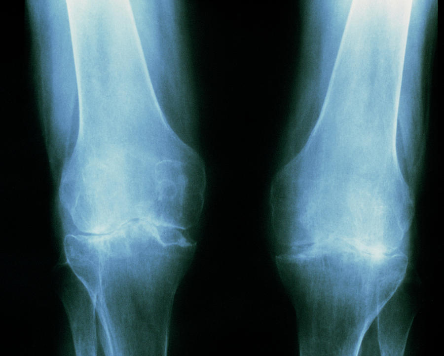
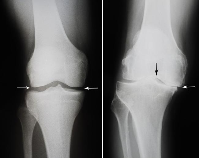
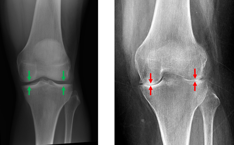
X Ray Knee Arthritis Osteoarthritis (oa) of the knee is very common and is a major cause of morbidity, especially in the older population. for a general discussion on osteoarthritis, please see the general osteoarthritis article. The four tell tale signs of osteoarthritis in the knee visible on an x ray include joint space narrowing, bone spurs, irregularity on the surface of the joints, and sub cortical cysts.

X Ray Knee Arthritis This article discusses what arthritis looks like on an x ray, offering insights into how this imaging technique is pivotal in identifying and managing arthritis. key signs of arthritis on an x ray x ray imaging provides a detailed picture of bone and joint health, crucial for diagnosing arthritis. Common findings of knee osteoarthritis from x rays. typically, the cartilage in one compartment of the joint (that is, the medial, lateral, or anterior patellofemoral joint compartment) is most severely affected. the standing x rays may show narrowing of the involved joint space of the knee. Getting an x ray of the knee is often a first step in diagnosing a knee condition. in many cases, knee x rays can help find the cause of pain, tenderness, or swelling. x rays are best at showing bone, but can also reveal soft tissue changes and signs of arthritis. Learn how x rays and imaging help diagnose knee arthritis. discover what joint space narrowing, bone spurs, and other signs mean for your treatment & mobility.

X Ray Knee Arthritis Getting an x ray of the knee is often a first step in diagnosing a knee condition. in many cases, knee x rays can help find the cause of pain, tenderness, or swelling. x rays are best at showing bone, but can also reveal soft tissue changes and signs of arthritis. Learn how x rays and imaging help diagnose knee arthritis. discover what joint space narrowing, bone spurs, and other signs mean for your treatment & mobility. Knee arthritis can affect one side of the joint more than the other. this x ray image shows how the cushioning cartilage has worn away, allowing bone to touch bone. Osteoarthritis results in characteristic x ray appearances including joint space narrowing, formation of osteophytes (bone spurs), articular surface cortical irregularity and or sclerosis, and formation of sub cortical cysts (geodes). Osteoarthritis of the knee is very common and is a major cause of morbidity, especially in the older population. To get pictures of the affected joint, your healthcare professional might recommend: x rays. cartilage doesn't show up on x ray images, but cartilage loss is revealed by a narrowing of the space between the bones in your joint. an x ray also can show bone spurs around a joint. magnetic resonance imaging (mri).

Comments are closed.