Diagram Of The Human Heart Blood Flow Through The Heart Pathways And Circulation Cardiac

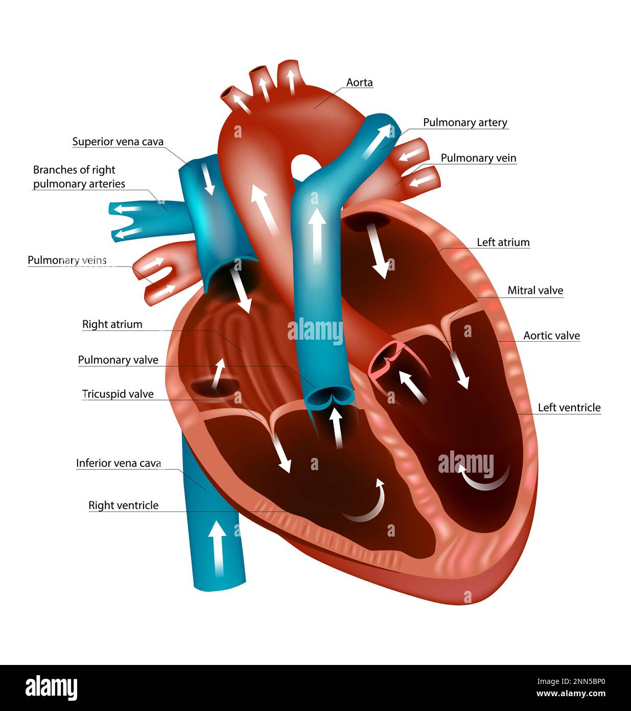
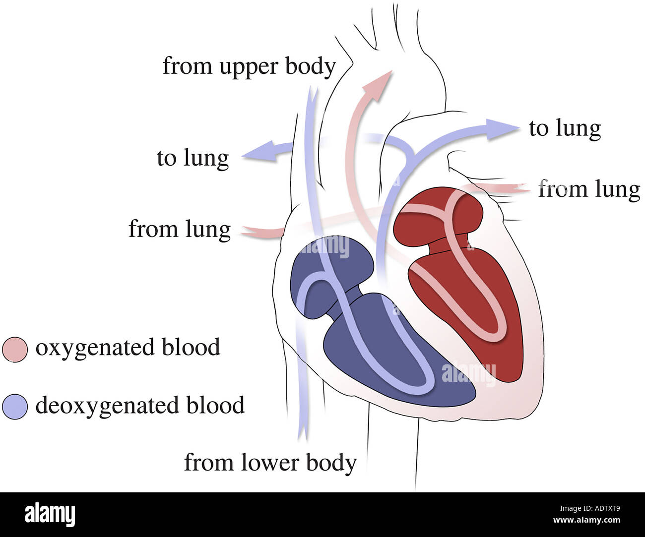
Diagram Of The Human Heart Blood Flow Through The Heart Pathways And The cardiac circulation process begins in the right atrium, where deoxygenated blood from the body enters through the venae cavae. this blood then flows through the tricuspid valve into the right ventricle, initiating the pulmonary circuit. A labeled heart diagram helps you understand the structure of human heart, which pumps blood through body. learn the structure and several heart conditions.

Diagram Of The Human Heart Blood Flow Through The Heart Pathways And The human heart serves as the body’s central pump, circulating blood continuously. this article explores the heart’s structure and function, detailing its physical layout, the pathway of blood, and its internal electrical system. Learn about the heart's anatomy, how it functions, blood flow through the heart and lungs, its location, artery appearance, and how it beats. Arteries and veins link your heart to the rest of the circulatory system. veins bring blood to your heart. arteries take blood away from your heart. your heart valves help control the direction the blood flows. heart valves control the flow of blood so that it moves in the right direction. the valves prevent blood from flowing backward. Visual representations such as the blood flow in heart diagram or the cardiac blood flow diagram are essential tools for learning. these diagrams clearly map the sequential flow through each chamber and valve, reinforcing the textual description of the blood’s path.

Heart Blood Flow Simple Anatomy Diagram Cardiac 41 Off Arteries and veins link your heart to the rest of the circulatory system. veins bring blood to your heart. arteries take blood away from your heart. your heart valves help control the direction the blood flows. heart valves control the flow of blood so that it moves in the right direction. the valves prevent blood from flowing backward. Visual representations such as the blood flow in heart diagram or the cardiac blood flow diagram are essential tools for learning. these diagrams clearly map the sequential flow through each chamber and valve, reinforcing the textual description of the blood’s path. Blood flow through the heart. blood flows through the heart, lungs, and body in a series of steps. after delivering oxygen and nutrients to all your organs and tissues, your blood enters your heart and flows to your lungs to gain oxygen and get rid of waste. This detailed anatomical diagram presents both external and internal views of the heart, showcasing the complex pathway of oxygenated and deoxygenated blood flow that sustains life. The diagram illustrating the blood flow through the heart shows the various chambers and valves that ensure efficient circulation. the heart consists of four chambers: two atria (left and right) and two ventricles (left and right). the atria receive deoxygenated blood, while the ventricles pump oxygenated blood out to the body. Explore a labeled diagram of the heart and learn about the flow of blood through its chambers and vessels. understand how the heart pumps oxygenated and deoxygenated blood to different parts of the body.

Blood Flow Through Heart Pathways Circulation Stock Vector Royalty Blood flow through the heart. blood flows through the heart, lungs, and body in a series of steps. after delivering oxygen and nutrients to all your organs and tissues, your blood enters your heart and flows to your lungs to gain oxygen and get rid of waste. This detailed anatomical diagram presents both external and internal views of the heart, showcasing the complex pathway of oxygenated and deoxygenated blood flow that sustains life. The diagram illustrating the blood flow through the heart shows the various chambers and valves that ensure efficient circulation. the heart consists of four chambers: two atria (left and right) and two ventricles (left and right). the atria receive deoxygenated blood, while the ventricles pump oxygenated blood out to the body. Explore a labeled diagram of the heart and learn about the flow of blood through its chambers and vessels. understand how the heart pumps oxygenated and deoxygenated blood to different parts of the body.

Comments are closed.