Intracranial Hemorrhage Pacs

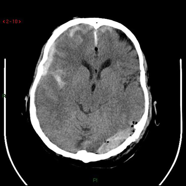
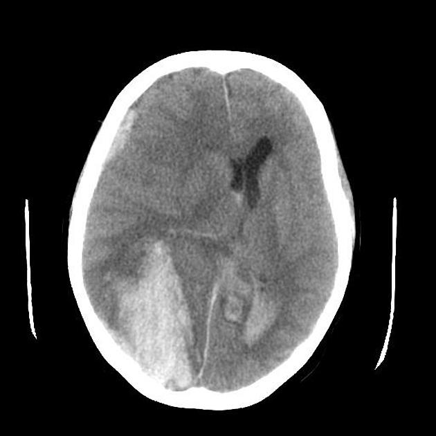
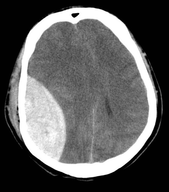
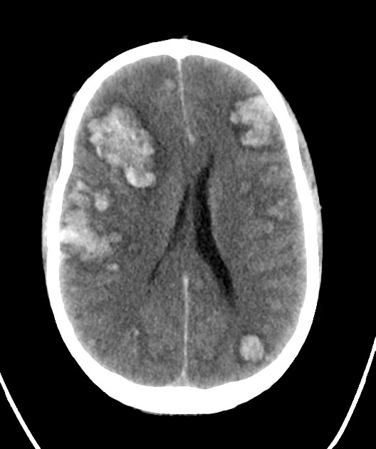
Intracranial Hemorrhage Pdf Neurology Nervous System An intracerebral hemorrhage, or intraparenchymal cerebral hemorrhage, is a subset of an intracranial hemorrhage and encompasses a number of entities that have in common the acute accumulation of blood within the parenchyma of the brain. A noncontrast head ct is obtained and the study is routed to a pacs for interpretation. concurrently, an algorithm evaluates the cross sectional imaging to screen for acute intracranial blood products, indicating present, absent, or undetermined.

Intracranial Hemorrhage Pacs Intracranial hemorrhage (ich) is a collective term encompassing many different conditions characterized by the extravascular accumulation of blood within different intracranial spaces. This guideline recommends development of regional systems that provide initial intracerebral hemorrhage (ich) care and the capacity, when appropriate, for rapid transfer to facilities with neurocritical care and neurosurgical capabilities. Fetal intracranial hemorrhage may occur either within the cerebral ventricles, subdural space or infratentorial fossa. hemorrhages can occur in a number of situations: the sonographic appearance of fetal intracranial hemorrhage is extremely variable, depending on its location and age of the hemorrhage. Viz hemorrhage harnesses the power of ai to provide precise and reliable detection and quantification of intracerebral hemorrhages.

Intracranial Hemorrhage Pacs Fetal intracranial hemorrhage may occur either within the cerebral ventricles, subdural space or infratentorial fossa. hemorrhages can occur in a number of situations: the sonographic appearance of fetal intracranial hemorrhage is extremely variable, depending on its location and age of the hemorrhage. Viz hemorrhage harnesses the power of ai to provide precise and reliable detection and quantification of intracerebral hemorrhages. Intracranial hemorrhage refers to bleeding within the intracranial cavity and is, therefore, a catch all term which includes parenchymal (intra axial) hemorrhage and the various types of extra axial hemorrhage including, subarachnoid, subdural and extradural hemorrhage. Intracranial hemorrhage: the daily pance blueprint a 44 year old woman presents to the emergency department with the sudden onset of the “worst headache of my life” while bending over to pick up a box. she reports associated nausea but no focal neurologic deficits. a non contrast head ct performed within 3 hours of symptom onset is negative. Cerebral hemorrhagic contusions are a type of intracerebral hemorrhage and are common in the setting of significant head injury. they are usually characterized on ct as hyperdense foci in the frontal lobes adjacent to the floor of the anterior cranial fossa and in the temporal poles. Intracranial hemorrhage (ich) and resulting neurological disability are severe complications for a subset of infants with severe hemophilia a (ha). while prophylactic factor replacement reduces bleeding risk, it is typically delayed until after age 1 due to risks associated with central venous access placement.

Intracranial Hemorrhage Pacs Intracranial hemorrhage refers to bleeding within the intracranial cavity and is, therefore, a catch all term which includes parenchymal (intra axial) hemorrhage and the various types of extra axial hemorrhage including, subarachnoid, subdural and extradural hemorrhage. Intracranial hemorrhage: the daily pance blueprint a 44 year old woman presents to the emergency department with the sudden onset of the “worst headache of my life” while bending over to pick up a box. she reports associated nausea but no focal neurologic deficits. a non contrast head ct performed within 3 hours of symptom onset is negative. Cerebral hemorrhagic contusions are a type of intracerebral hemorrhage and are common in the setting of significant head injury. they are usually characterized on ct as hyperdense foci in the frontal lobes adjacent to the floor of the anterior cranial fossa and in the temporal poles. Intracranial hemorrhage (ich) and resulting neurological disability are severe complications for a subset of infants with severe hemophilia a (ha). while prophylactic factor replacement reduces bleeding risk, it is typically delayed until after age 1 due to risks associated with central venous access placement.

Intracranial Hemorrhage Pacs Cerebral hemorrhagic contusions are a type of intracerebral hemorrhage and are common in the setting of significant head injury. they are usually characterized on ct as hyperdense foci in the frontal lobes adjacent to the floor of the anterior cranial fossa and in the temporal poles. Intracranial hemorrhage (ich) and resulting neurological disability are severe complications for a subset of infants with severe hemophilia a (ha). while prophylactic factor replacement reduces bleeding risk, it is typically delayed until after age 1 due to risks associated with central venous access placement.

Comments are closed.