Middle Cerebral Artery Anatomy Radiology Notes Vrogue Co

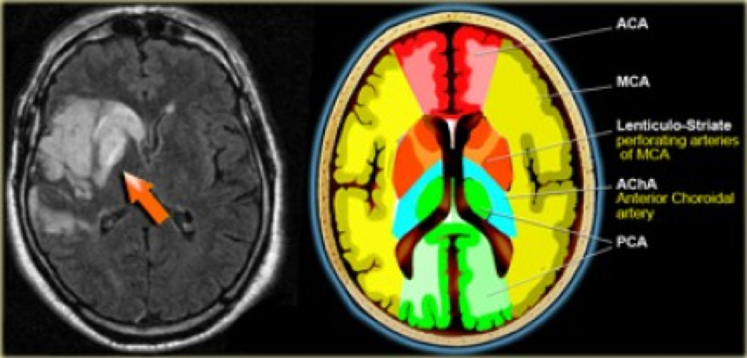
Middle Cerebral Artery Anatomy Radiology Notes Vrogue Co The mca arises from the internal carotid artery as the larger of the two main terminal branches (the other being the anterior cerebral artery), coursing laterally into the lateral sulcus where it branches to perfuse the cerebral cortex. The middle cerebral artery (mca) is, along with the anterior cerebral artery, one of the terminal branches of the internal carotid artery. it is responsible for supplying blood to large parts of the cerebral hemispheres.

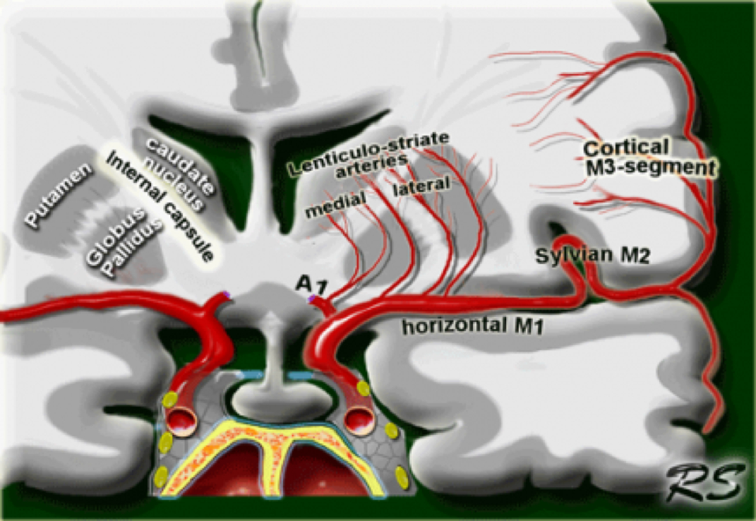
Middle Cerebral Artery Anatomy Radiology Notes Vrogue Co The anterior commissure is a bundle of white fibers that connects the two cerebral hemispheres across the middle line. at this level frequently perivascular csf spaces of virchow robin are seen. The middle cerebral artery (mca) is one of the three major paired arteries that supply blood to the brain. Advances in neuroimaging and endovascular therapy have further highlighted the clinical importance of this vessel, enabling targeted interventions for acute stroke management. this activity explores the mca’s anatomy, functional role, and clinical implications. Anatomically, the mca is divided into two segments (m1 and m2)(3). however, in radiology and surgery, the middle cerebral artery is divided into four parts (m1, m2, m3, and m4)(4). this segment is also called the horizontal segment or the sphenoid part of the mca.

Middle Cerebral Artery Anatomy Radiology Notes Advances in neuroimaging and endovascular therapy have further highlighted the clinical importance of this vessel, enabling targeted interventions for acute stroke management. this activity explores the mca’s anatomy, functional role, and clinical implications. Anatomically, the mca is divided into two segments (m1 and m2)(3). however, in radiology and surgery, the middle cerebral artery is divided into four parts (m1, m2, m3, and m4)(4). this segment is also called the horizontal segment or the sphenoid part of the mca. One of the three major paired arteries that supply blood the brain. [foogallery id=”7185″] m1 – sphenoidal or horizontal segment m2 – insular segment m3…. This five part educational series aims to incorporate extensive annotated digital subtraction angiography (dsa) images to review the anatomy of the anterior and posterior circulation most critical to the neurointerventionalist. Annotated image of part of the circle of willis showing the middle cerebral arteries. the internal carotid artery coming up from the neck divides into the middle and anterior cerebral arteries. For these reasons, we present this anatomic review of the mca, reviewing its segments and anatomic limits, its branching patterns, and its anatomic variants.

Middle Cerebral Artery Anatomy Radiology Notes One of the three major paired arteries that supply blood the brain. [foogallery id=”7185″] m1 – sphenoidal or horizontal segment m2 – insular segment m3…. This five part educational series aims to incorporate extensive annotated digital subtraction angiography (dsa) images to review the anatomy of the anterior and posterior circulation most critical to the neurointerventionalist. Annotated image of part of the circle of willis showing the middle cerebral arteries. the internal carotid artery coming up from the neck divides into the middle and anterior cerebral arteries. For these reasons, we present this anatomic review of the mca, reviewing its segments and anatomic limits, its branching patterns, and its anatomic variants.

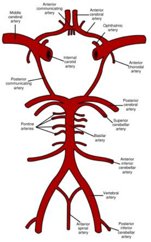
Middle Cerebral Artery Anatomy Radiology Notes Annotated image of part of the circle of willis showing the middle cerebral arteries. the internal carotid artery coming up from the neck divides into the middle and anterior cerebral arteries. For these reasons, we present this anatomic review of the mca, reviewing its segments and anatomic limits, its branching patterns, and its anatomic variants.

Comments are closed.