

Small Bowel Radiograph Diagram | Quizlet
Small Bowel Radiograph Diagram | Quizlet Radiography is an imaging technique using x rays, gamma rays, or similar ionizing radiation and non ionizing radiation to view the internal form of an object. applications of radiography include medical ("diagnostic" radiography and "therapeutic radiography") and industrial radiography. A radiograph (or plain radiograph although the word 'plain' is strictly superfluous) is the radiologist's preferred term for the static image generated following the passage of x rays through the patient.

PA 30 Minutes Small Bowel Radiograph - Slide 7 Diagram | Quizlet
PA 30 Minutes Small Bowel Radiograph - Slide 7 Diagram | Quizlet Medical radiography is a broad term that covers several types of studies that require the visualization of the internal parts of the body using x ray techniques. X rays, or plain radiographs, are a way for providers to get pictures of the inside of your body. x rays use radiation to create black and white images that a radiologist reads. x rays are most commonly used to look at bones and joints, but providers can use them to quickly diagnose other conditions, like lung infections, too. Radiography is a crucial imaging technique that plays a vital role in both healthcare and industrial fields. it uses ionizing radiation, such as x rays or gamma rays, to capture images of the internal structures of the body or objects. The meaning of radiograph is a picture produced on a sensitive surface by a form of radiation other than visible light; specifically : an x ray or gamma ray photograph.

Radiograph Showing Small Bowel Obstruction – GPnotebook
Radiograph Showing Small Bowel Obstruction – GPnotebook Radiography is a crucial imaging technique that plays a vital role in both healthcare and industrial fields. it uses ionizing radiation, such as x rays or gamma rays, to capture images of the internal structures of the body or objects. The meaning of radiograph is a picture produced on a sensitive surface by a form of radiation other than visible light; specifically : an x ray or gamma ray photograph. Some x ray exams may use an iodine based contrast material or barium to help improve the visibility of specific organs, blood vessels, tissues or bone. for the benefits and risks of a specific x ray procedure, how to prepare, and more, select a topic below. x ray tests, treatments and procedures. For patients with high clinical suspicion, plain radiographs are indicated for initial evaluation of focal bone pain. parasitic lesions were diagnosed by imaging of characteristic space occupying lesions in abdominal organs (ultrasound) or the lungs (radiographs). Radiography is the most readily available imaging method. typically, it is the first imaging method indicated to evaluate the extremities, chest, and sometimes the spine and abdomen. these areas contain important structures with densities that differ from those of adjacent tissues. Radiography is the art and science of using radiation to provide images of the tissues, organs, bones, and vessels that comprise the human body. radiologists, physicians who have had special training in interpreting diagnostic images, read or diagnose these images.

Radiograph Showing Small Bowel Obstruction | Download Scientific Diagram
Radiograph Showing Small Bowel Obstruction | Download Scientific Diagram Some x ray exams may use an iodine based contrast material or barium to help improve the visibility of specific organs, blood vessels, tissues or bone. for the benefits and risks of a specific x ray procedure, how to prepare, and more, select a topic below. x ray tests, treatments and procedures. For patients with high clinical suspicion, plain radiographs are indicated for initial evaluation of focal bone pain. parasitic lesions were diagnosed by imaging of characteristic space occupying lesions in abdominal organs (ultrasound) or the lungs (radiographs). Radiography is the most readily available imaging method. typically, it is the first imaging method indicated to evaluate the extremities, chest, and sometimes the spine and abdomen. these areas contain important structures with densities that differ from those of adjacent tissues. Radiography is the art and science of using radiation to provide images of the tissues, organs, bones, and vessels that comprise the human body. radiologists, physicians who have had special training in interpreting diagnostic images, read or diagnose these images.
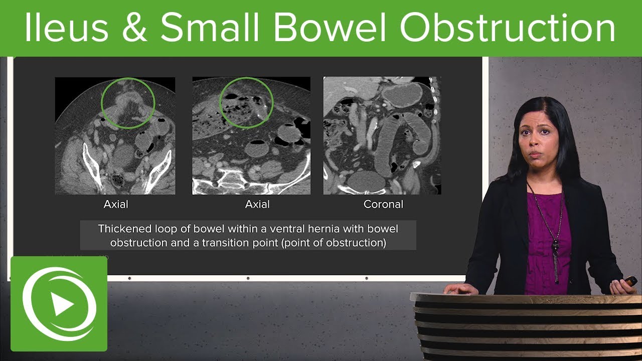
Bowel Obstruction and Ileus: Ileus & Small Bowel Obstruction – Radiology | Lecturio
Bowel Obstruction and Ileus: Ileus & Small Bowel Obstruction – Radiology | Lecturio
Related image with radiograph showing small bowel obstruction download scientific diagram


Related image with radiograph showing small bowel obstruction download scientific diagram


About "Radiograph Showing Small Bowel Obstruction Download Scientific Diagram"
Comments are closed.