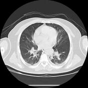
Saddle Pulmonary Embolism Echo Clip

Saddle Pulmonary Embolism A Rare Case Of Proximal Pulmonary Embolism #pe #echo. Saddle pulmonary embolism seen on echo. this astute finding by the echo tech allowed for early diagnosis and treatment.

Saddle Pulmonary Embolism Radiology Reference Article Radiopaedia Org Saddle pulmonary embolism commonly refers to a large pulmonary embolism that straddles the bifurcation of the pulmonary trunk, extending into the left and right pulmonary arteries. if large enough, it can completely obstruct both left and right pulmonary arteries resulting in right heart failure and, unless treatment is prompt, death. Surgical videopulmonary embolectomy with retrograde pulmonary perfusion for saddle pulmonary embolus. specialty: cardiothoracic surgery. subspecialty: thoracic surgery. society of thoracic surgeons. | published: 02 2021. ** this video may contain materials that have been removed due to copyright restrictions. translation disclaimer. 0comments. Computed tomography pulmonary angiogram showing a large saddle pulmonary embolus with extensive bilateral clot burden (arrowheads). the main pulmonary artery and ascending aorta diameters are roughly equivalent, indicative of pulmonary. We present a rare case of an intermediate high risk saddle pe with extensive bilateral clot burden and evidence of right heart dysfunction, masquerading as micturition syncope and discovered by point of care echocardiography.

Saddle Pulmonary Embolism Everything You Need To Know About It Computed tomography pulmonary angiogram showing a large saddle pulmonary embolus with extensive bilateral clot burden (arrowheads). the main pulmonary artery and ascending aorta diameters are roughly equivalent, indicative of pulmonary. We present a rare case of an intermediate high risk saddle pe with extensive bilateral clot burden and evidence of right heart dysfunction, masquerading as micturition syncope and discovered by point of care echocardiography. This is an uncommon example of central pulmonary embolism which can be suspected on non contrast ct. a saddle pulmonary embolism (spe) occurs when a blood clot lodges at the bifurcation of the main pulmonary artery, extending into both the right and left pulmonary arteries. Computed tomographic pulmonary angiography showed a large saddle pulmonary embolus involving the main, left, and right pulmonary arteries (c, arrow), which correlated with the tte findings. thrombolytic therapy and anticoagulation were initiated. the patient had an uneventful hospital course. A saddle pulmonary embolism is when a large blood clot gets stuck in the main pulmonary artery. it’s a type of pulmonary embolism (pe), which is a blockage in one of the arteries of the lungs. We describe a case of a saddle embolus of the main pulmonary artery visualized by real time three dimensional echocardiography and successfully treated with intravenous unfractionated heparin, followed by oral anticoagulation achieving a complete dissolution of the thrombus.

Comments are closed.