Stock Brain Hemorrhages Illustrated Verdict

Stock Brain Hemorrhages Illustrated Verdict Types of hemorrhages on surface and in coverings of brain: caput succadeaneaum, cephalhematoma, subgaleal hematoma, and subdural hematoma. 1. normal anatomy of surface of brain and coverings. 2. subdural bleed. 3. subarachnoid bleed. 1. normal anatomy of surface of brain and coverings. 2. progression of subdural hematoma. 1. Identify signs and symptoms indicative of intracranial hemorrhage in patients of varying age groups and demographics. implement evidence based protocols and proper evaluation, imaging, and monitoring protocols for timely diagnosis and management of intracranial hemorrhage.

Stock Brain Hemorrhages Illustrated Verdict Patients with intracerebral hemorrhage present with focal neurologic signs that are abrupt in onset but not instantaneous, as occurs with embolic ischemic stroke. There are two types of brain bleeds that occur inside the brain tissue itself: intracerebral hemorrhage: this bleeding occurs in the lobes, brainstem and cerebellum of your brain. this is bleeding anywhere within the brain tissue itself. Intracranial hemorrhage is a collective term encompassing many different conditions characterized by the extravascular accumulation of blood within different intracranial spaces. a simple categorization is based on location:. Amphora parallel wiki koen · datasets at hugging facetrain · 30.2k rows.

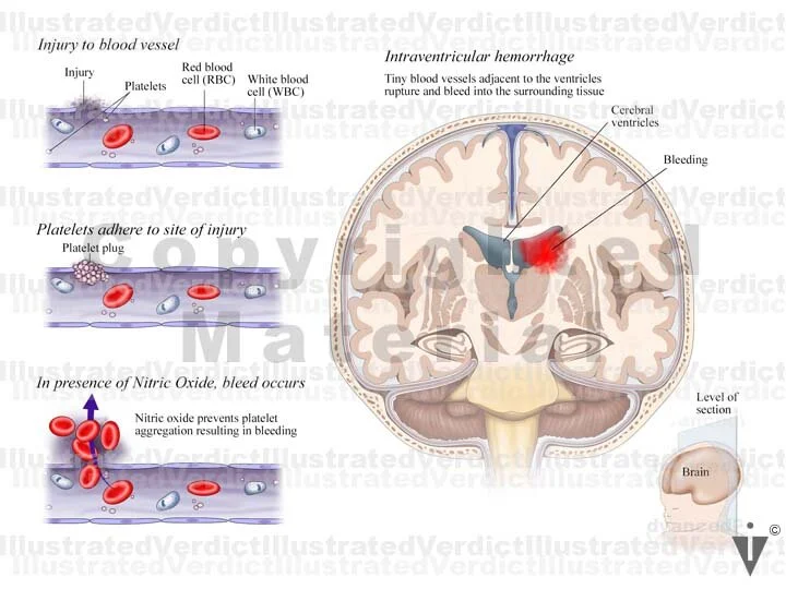
Stock Brain Hemorrhages Illustrated Verdict Intracranial hemorrhage is a collective term encompassing many different conditions characterized by the extravascular accumulation of blood within different intracranial spaces. a simple categorization is based on location:. Amphora parallel wiki koen · datasets at hugging facetrain · 30.2k rows. Intracerebral hemorrhage (ich) is a severe clinical emergency caused by bleeding into brain parenchyma. currently, there are no effective treatments to improve ich outcomes. developing new therapies for ich relies on a thorough understanding of ich pathophysiology and good in vitro models that enable mechanistic research. Ultra early blood pressure reduction attenuates hematoma growth and improves outcome in intracerebral hemorrhage. ann neurol. 2020;88:388–395. doi: 10.1002 ana.25793. 5. Focal neurologic deficits with intracerebral hemorrhage in the cerebral hemispheres (lobar hemorrhage) correspond to the location of the hemorrhage and its transection of white matter tracts;. 5. subdural hematoma board 11 1. axial view of brain w subarachnoid hemorrhages 2. normal meninges 3. subarachnoid hemorrhage board 12 1 3. embolic stroke 4 6. hemorrhagic stroke board 13 1. fetal brain at 26 30 weeks 2. germinal matrix hemorrhage board 14 1. normal anatomy: coronal view of brain 2. unilateral periventricular intraventricular.

Stock Brain Hemorrhages Illustrated Verdict Intracerebral hemorrhage (ich) is a severe clinical emergency caused by bleeding into brain parenchyma. currently, there are no effective treatments to improve ich outcomes. developing new therapies for ich relies on a thorough understanding of ich pathophysiology and good in vitro models that enable mechanistic research. Ultra early blood pressure reduction attenuates hematoma growth and improves outcome in intracerebral hemorrhage. ann neurol. 2020;88:388–395. doi: 10.1002 ana.25793. 5. Focal neurologic deficits with intracerebral hemorrhage in the cerebral hemispheres (lobar hemorrhage) correspond to the location of the hemorrhage and its transection of white matter tracts;. 5. subdural hematoma board 11 1. axial view of brain w subarachnoid hemorrhages 2. normal meninges 3. subarachnoid hemorrhage board 12 1 3. embolic stroke 4 6. hemorrhagic stroke board 13 1. fetal brain at 26 30 weeks 2. germinal matrix hemorrhage board 14 1. normal anatomy: coronal view of brain 2. unilateral periventricular intraventricular.

Stock Brain Hemorrhages Illustrated Verdict Focal neurologic deficits with intracerebral hemorrhage in the cerebral hemispheres (lobar hemorrhage) correspond to the location of the hemorrhage and its transection of white matter tracts;. 5. subdural hematoma board 11 1. axial view of brain w subarachnoid hemorrhages 2. normal meninges 3. subarachnoid hemorrhage board 12 1 3. embolic stroke 4 6. hemorrhagic stroke board 13 1. fetal brain at 26 30 weeks 2. germinal matrix hemorrhage board 14 1. normal anatomy: coronal view of brain 2. unilateral periventricular intraventricular.

Comments are closed.