Thoracic Aortic Aneurysms Radiology Key
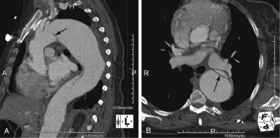
Thoracic Aortic Aneurysms Radiology Key Thoracic aortic aneurysms are less common than abdominal aneurysms, and the prevalence depends on the etiology (fig. 92 1). of note, aneurysms due to systemic arterial disease have less male preponderance and a more advanced age at presentation than abdominal aneurysms do, resulting in greater comorbid disease. 2. Thoracic aortic aneurysm (taa) is a chronic condition that manifests as progressive dilation of the thoracic aorta resulting from degradation of the normal smooth muscle cells and extracellular matrix proteins that provide integrity to the aortic wall.
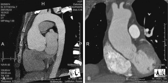
Thoracic Aortic Aneurysms Radiology Key Aneurysms—to provide a durable conduit for aortic blood flow across the entire longitudinal extent of the aneurysm, resulting in aneurysm sac depressurization, thrombosis formation, and eventual stabilization or regression in size. Terminology. the normal aortic diameter varies based on age, sex, and body surface area. in general, the term aneurysm is used when the axial diameter is >5.0 cm for the ascending aorta and >4.0 cm for the descending aorta 12 when enlarged above normal but not reaching aneurysmal definition, the terms dilatation ectasia can be used 9,12 epidemiology. This review will summarize the imaging evaluation and underlying pathology relevant to the diagnosis of thoracic aortic aneurysm. key points: cross sectional imaging (cta and mra) plays a central role in management of patients with thoracic aortic aneurysm. maximal aortic diameter is the primary metric used to estimate risk and determine the. Computed tomography angiogra phy (cta) and magnetic resonance angiogra phy (mra) are key to characterizing the an eurysm and the rest of the vasculature, while ultrasonography or echocardiography assist in assessment and surveillance, and catheter angiography is the gold standard for renal and splenic aneurysm.
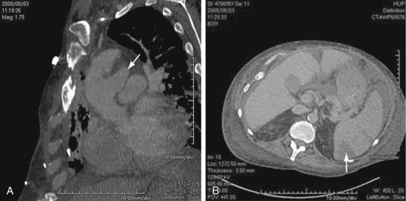
Thoracic Aortic Aneurysms Radiology Key This review will summarize the imaging evaluation and underlying pathology relevant to the diagnosis of thoracic aortic aneurysm. key points: cross sectional imaging (cta and mra) plays a central role in management of patients with thoracic aortic aneurysm. maximal aortic diameter is the primary metric used to estimate risk and determine the. Computed tomography angiogra phy (cta) and magnetic resonance angiogra phy (mra) are key to characterizing the an eurysm and the rest of the vasculature, while ultrasonography or echocardiography assist in assessment and surveillance, and catheter angiography is the gold standard for renal and splenic aneurysm. Aortic aneurysms can be categorized based on location into thoracic (taa, inccidence 0.16%) and abdominal (aaa, incidence 4%) 1. the prevalence of taa by location is 77% ascending, 5% arch, 10% descending and 8% thoracoabdominal 2. a retrospective analysis of 1,326 patients with taas noted a mean aortic diameter of 52 mm at time of diagnosis. Thoracic aortic aneurysm (taa) is a chronic condi tion that manifests as progressive dilation of the thoracic aorta resulting from degradation of the normal smooth muscle cells and extracellular ma trix proteins that provide integrity to the aortic wall. Thoracic aortic aneurysm (taa) is a chronic condition that manifests as progressive dilation of the thoracic aorta resulting from degradation of the normal smooth muscle cells and extracellular matrix proteins that provide integrity to the aortic wall. Our exhibit will review 3d imaging of the aorta as well as proper measurement and diagnosis of thoracic aortic aneurysms based upon the current guidelines.
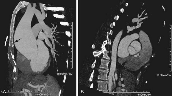
Thoracic Aortic Aneurysms Radiology Key Aortic aneurysms can be categorized based on location into thoracic (taa, inccidence 0.16%) and abdominal (aaa, incidence 4%) 1. the prevalence of taa by location is 77% ascending, 5% arch, 10% descending and 8% thoracoabdominal 2. a retrospective analysis of 1,326 patients with taas noted a mean aortic diameter of 52 mm at time of diagnosis. Thoracic aortic aneurysm (taa) is a chronic condi tion that manifests as progressive dilation of the thoracic aorta resulting from degradation of the normal smooth muscle cells and extracellular ma trix proteins that provide integrity to the aortic wall. Thoracic aortic aneurysm (taa) is a chronic condition that manifests as progressive dilation of the thoracic aorta resulting from degradation of the normal smooth muscle cells and extracellular matrix proteins that provide integrity to the aortic wall. Our exhibit will review 3d imaging of the aorta as well as proper measurement and diagnosis of thoracic aortic aneurysms based upon the current guidelines.
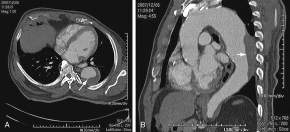
Thoracic Aortic Aneurysms Radiology Key Thoracic aortic aneurysm (taa) is a chronic condition that manifests as progressive dilation of the thoracic aorta resulting from degradation of the normal smooth muscle cells and extracellular matrix proteins that provide integrity to the aortic wall. Our exhibit will review 3d imaging of the aorta as well as proper measurement and diagnosis of thoracic aortic aneurysms based upon the current guidelines.
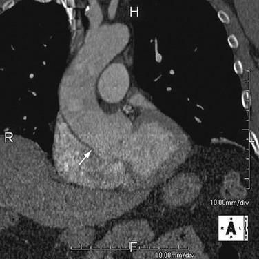
Thoracic Aortic Aneurysms Radiology Key

Comments are closed.