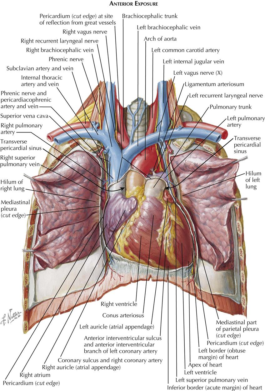
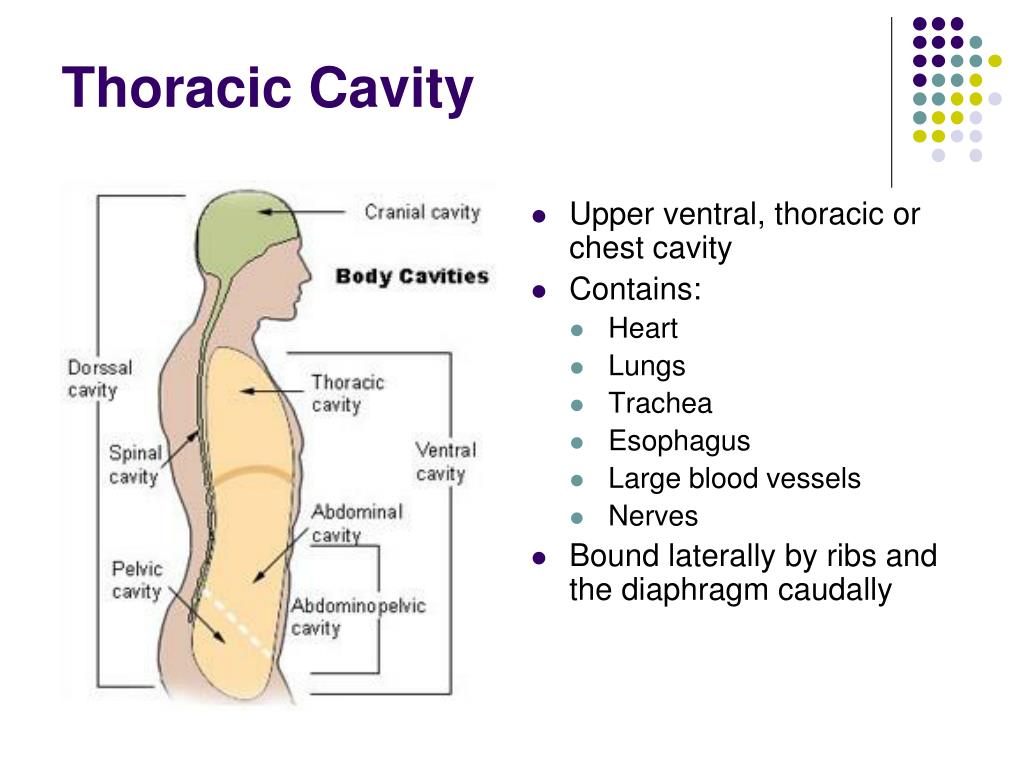
Thoracic Region Anatomy Every Chest Cavity Structure Labeled

Thoracic Cavity Labeled Thoracic region anatomy every chest cavity structure labeled! 🫁 i use axis scientific models from anatomy warehouse in my videos. links below!torso model (with all the organs):. The thoracic, or chest wall, consists of a skeletal framework, fascia, muscles, and neurovasculature – all connected together to form a strong and protective yet flexible cage. the thorax has two major openings: the superior thoracic aperture found superiorly and the inferior thoracic aperture located inferiorly.

Thoracic Cavity Model Labeled In this section, learn more about the areas of the thorax, bones of the thorax, muscles of the thorax, organs of the thorax and the vasculature of the thorax. explore the anatomy of the human thorax. this comprehensive guide covers the thoracic cavity's vital structures and their functions. learn more here. Arteries of the thoracic cavity. the arch of the aorta has three major branches: the brachiocephalic trunk, left common carotid artery, and left subclavian artery. after the aortic arch, the aorta begins its descent, becoming the thoracic aorta at the level of the sternal angle and the abdominal aorta once it passes through the aortic hiatus in. The thorax or the chest (a.k.a. the upper torso) is part of the human body which is located between the neck (above) and the abdomen (below). the thorax includes an outer thoracic wall and an inner thoracic cavity: thoracic wall: the bony framework of the thoracic wall is formed by the sternum (in front) and twelve thoracic vertebrae (at the. Your thoracic cavity is a space in your chest that contains organs, blood vessels, nerves and other important body structures. it’s divided into three main parts: right pleural cavity, left pleural cavity and mediastinum. the five organs in your thoracic cavity are your heart, lungs, esophagus, trachea and thymus.

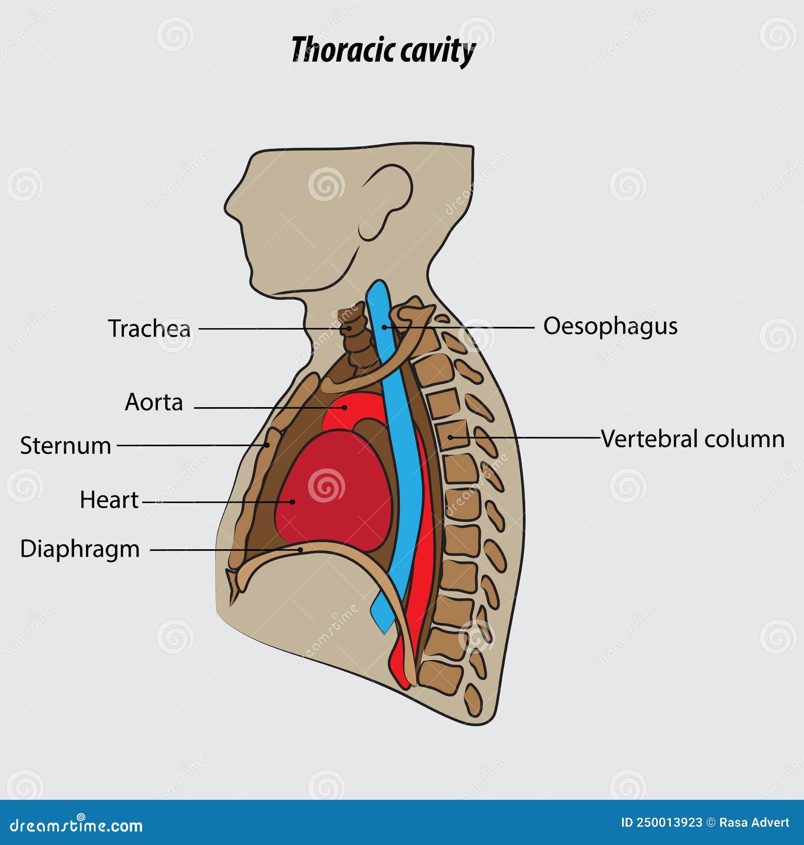
Thoracic Cavity Vector Illustration Drawing Labeled Stock Vector The thorax or the chest (a.k.a. the upper torso) is part of the human body which is located between the neck (above) and the abdomen (below). the thorax includes an outer thoracic wall and an inner thoracic cavity: thoracic wall: the bony framework of the thoracic wall is formed by the sternum (in front) and twelve thoracic vertebrae (at the. Your thoracic cavity is a space in your chest that contains organs, blood vessels, nerves and other important body structures. it’s divided into three main parts: right pleural cavity, left pleural cavity and mediastinum. the five organs in your thoracic cavity are your heart, lungs, esophagus, trachea and thymus. • the skeleton of the thoracic wall consists of 12 thoracic vertebrae, 12 pairs of ribs and costal cartilages and the sternum. • thoracic cavity – roofed in above lung apices by suprapleural membrane and floored by. 3d interactive modules and video tutorials on the anatomy of the thoracic cavity, including the heart, lungs, breast, chest wall, and respiratory tract. The thorax, or chest, is a complex structure that houses vital organs, provides support and protection, and enables respiration. this section of the 3d anatomy atlas explores the thoracic region, highlighting its skeletal framework, muscular system, vascular network, nerves, and internal organs. Start studying anatomy chapter 1: labeling thoracic cavity. learn vocabulary, terms, and more with flashcards, games, and other study tools.

Structure Of Thoracic Cavity Diagram Quizlet • the skeleton of the thoracic wall consists of 12 thoracic vertebrae, 12 pairs of ribs and costal cartilages and the sternum. • thoracic cavity – roofed in above lung apices by suprapleural membrane and floored by. 3d interactive modules and video tutorials on the anatomy of the thoracic cavity, including the heart, lungs, breast, chest wall, and respiratory tract. The thorax, or chest, is a complex structure that houses vital organs, provides support and protection, and enables respiration. this section of the 3d anatomy atlas explores the thoracic region, highlighting its skeletal framework, muscular system, vascular network, nerves, and internal organs. Start studying anatomy chapter 1: labeling thoracic cavity. learn vocabulary, terms, and more with flashcards, games, and other study tools.

Anatomy Of The Thoracic Chest Cavity Medical Art Works The thorax, or chest, is a complex structure that houses vital organs, provides support and protection, and enables respiration. this section of the 3d anatomy atlas explores the thoracic region, highlighting its skeletal framework, muscular system, vascular network, nerves, and internal organs. Start studying anatomy chapter 1: labeling thoracic cavity. learn vocabulary, terms, and more with flashcards, games, and other study tools.

Thoracic Cavity Labster

Comments are closed.