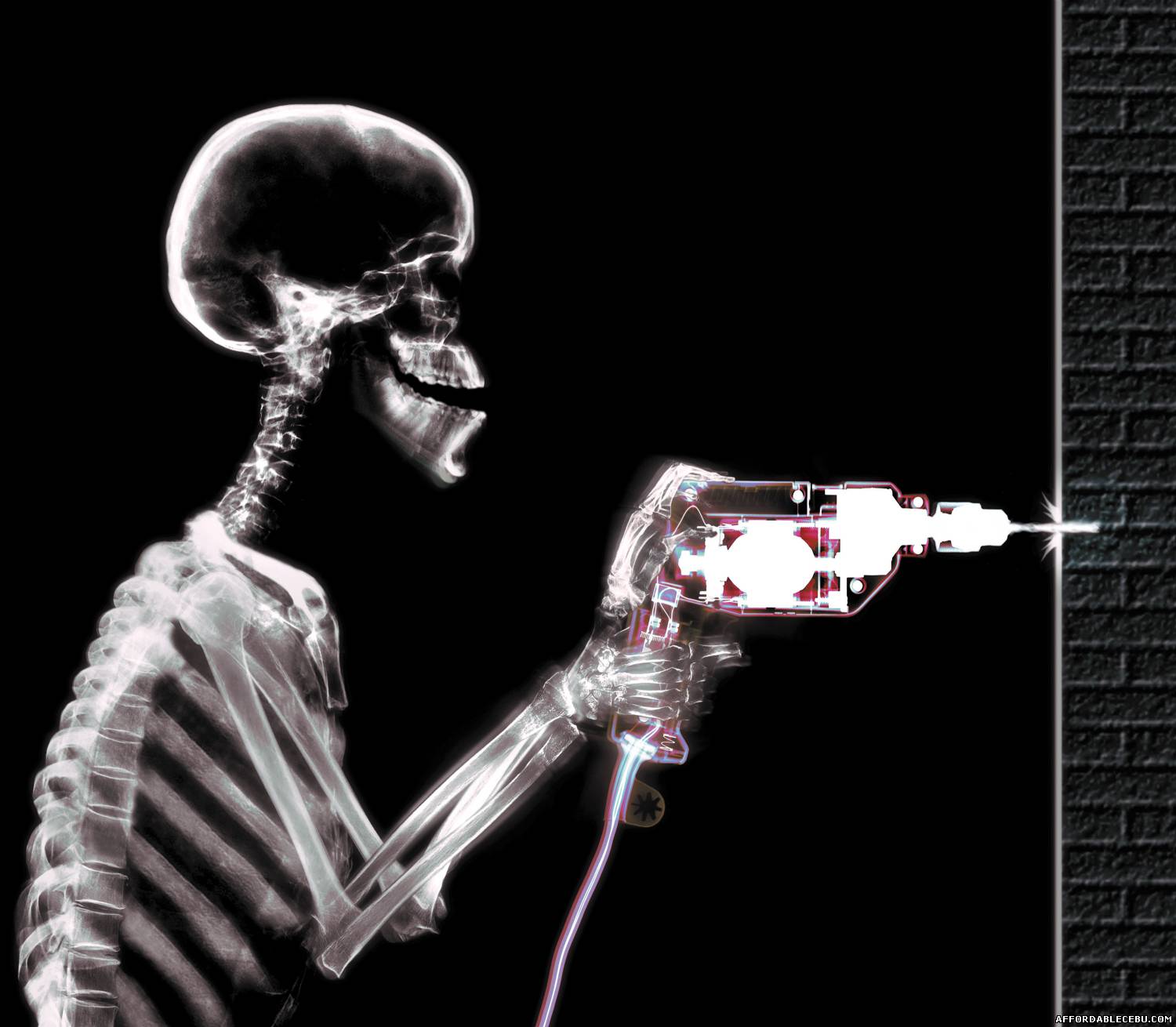
X Ray 2 0 Inside The Human Body In Incredible Color Anthony And Phil Butler Tedxchristchurch

Incredible Human X Ray Pictures Photoshop Graphics 626 When we think of an x ray, most of us visualize the classic, black and white image of bones a type of picture that hasn't changed much since the x ray's invention over 120 years ago. but,. But, leveraging technology from cern and the large hadron collider, phil and anthony butler a father and son team have brought the humble x ray into the modern world: full color, incredible resolution, and the ability to individually distinguish almost any type of material on the planet.

X Ray Of A Body X Ray Of Human Body Stock Illustration Illustration In this 2018 our changing world podcast from radio nz, professor anthony butler talks about the development of the revolutionary spectral molecular scanner that provides 3d colour images of objects inside the body, such as bone, soft tissue and artificial joints. The team — led by the father son duo of professors phil and anthony butler — shows off its latest mars scanners, which are capable of creating color 3d images of the inside of the body. When x rays travel through your body, they're absorbed by denser materials (bones) and pass right through softer ones (muscles and other tissues). the x rays that pass through unimpeded hit a. This colour x ray imaging technique could produce clearer and more accurate pictures and help doctors give their patients more accurate diagnoses. this is now a reality, thanks to a new zealand company that scanned, for the first time, a human body using a breakthrough colour medical scanner based on the medipix3 technology developed at cern.

X Ray Image Of Human Body Stock Illustration Adobe Stock When x rays travel through your body, they're absorbed by denser materials (bones) and pass right through softer ones (muscles and other tissues). the x rays that pass through unimpeded hit a. This colour x ray imaging technique could produce clearer and more accurate pictures and help doctors give their patients more accurate diagnoses. this is now a reality, thanks to a new zealand company that scanned, for the first time, a human body using a breakthrough colour medical scanner based on the medipix3 technology developed at cern. A new zealand company has generated the first 3d color x ray images of the human body by using an advanced medical scanner. the scanner utilizes cern's medipix3 technology and has been in. Medipix technology allows for a colorful look inside the human body. the colors above represent different energy levels of x ray photons recorded by the medipix technology. Creators of mars spectral x ray scanner, phil butler (right) and anthony butler. (source: university of canterbury) photon counting leads to the construction of the necessary spectrum, which can then be converted into the color images. therefore, it can be said that mars is, in fact, a form of ct. A new medical imaging device uses technology developed by particle physicists to produce full color, 3d images of the human body.

Comments are closed.